

On this page
From Guesswork to Precision
For generations, dentistry has relied on a form of sophisticated estimation. We are tasked with treating complex, three-dimensional anatomy—curved roots, variable bone widths, and hidden nerve canals—yet for decades, our only maps were flat, two-dimensional images. While Intraoral Periapical Radiographs (IOPAs) and Orthopantomograms (OPGs) are indispensable for basic screening, they fundamentally force the clinician to “interpret” reality rather than see it. We look at a shadow on a film and mentally reconstruct what the bone might look like, often hoping our estimation holds true once the flap is raised.
In the era of precision dentistry, this “guesswork” is becoming a significant clinical liability. In modern implantology and microscopic endodontics, not knowing the exact buccolingual width of a ridge or the precise curvature of a mesiobuccal root is no longer just a challenge; it is a risk—both to the patient’s safety and the clinician’s reputation.
The primary benefits of CBCT (Cone Beam Computed Tomography) lie in removing this uncertainty. It transitions the diagnostic process from interpretation to visualization. It allows us to peel back layers of tissue virtually before we ever pick up a scalpel. This guide explores how 3D imaging has transitioned from a luxury for elite centers to a medicolegal and clinical necessity for the everyday Indian practice, empowering you to plan treatments with absolute confidence rather than educated guesses.
The Dimension Gap: CBCT vs 2D X-ray
The fundamental limitation of traditional radiography is simple geometry: it forces a three-dimensional object onto a two-dimensional plane. In this compression, vital information is lost. When analyzing CBCT vs 2D xray, the most critical difference is the elimination of superimposition. On a standard periapical radiograph, the buccal and lingual cortical plates are smashed together into a single layer. A periapical lesion might be entirely obscured by a thick cortical plate, or a multi-rooted premolar might appear to have fused roots simply because they are overlapping in the 2D shadow.
Furthermore, 2D imaging is plagued by distortion and magnification. An OPG (Orthopantomogram) typically has a magnification factor ranging from 15% to 30%, which is often non-uniform. Relying on an OPG to measure vertical bone height for an implant is, therefore, a calculated risk; you are measuring a magnified shadow, not the bone itself.
CBCT technology solves these issues by utilizing isotropic voxels. Unlike the pixels in a 2D image which have only height and width, a voxel is a cube with equal dimensions in all three axes (height, width, and depth). This ensures a 1:1 measurement ratio. If you measure a distance of 12.5mm on your CBCT software, the anatomy is exactly 12.5mm in reality. This accuracy allows you to visualize the “third dimension”—the buccolingual width—ensuring that you are not just seeing the height of the bone, but its volume, quality, and exact proximity to vital structures.
Clinical Protocols: When to Use 3D Imaging


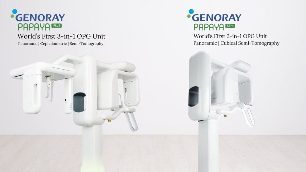 Dental OPG – GENORAY PAPAYA CUST
 Dental CBCT Machine – Genoray PAPAYA 3D PLUS
 Dental CBCT Machine – GENORAY PAPAYA 3D PREMIUM PLUS
 Genoray Port X-II
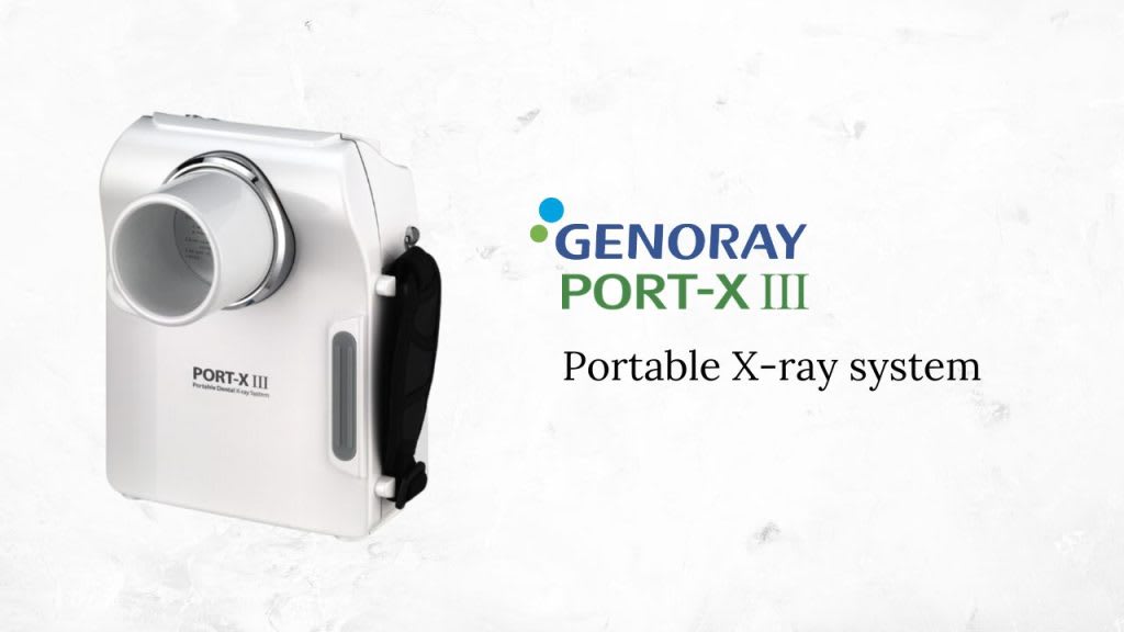 Genoray Port X-III
 Runyes Portable X-ray
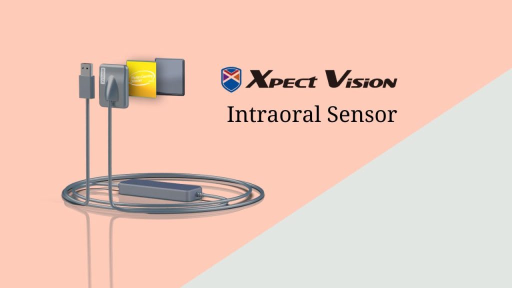 Xpect Vision Intraoral Sensor
 Carestream RVG CS 5200
 Gnatus Timex 70 E AC X-Ray Machine
Disclaimer
This article is for general information. Product specifications and features change with the manufacturer — confirm details with our team before you buy. Full disclaimer